August 4, 2022
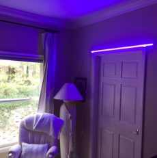
Salmonella Is Killed By SAFE Blue Light

HAS THE 405NM LIGHT BEEN TESTED TO KILL COMMON PATHOGENIC BACTERIA?
1. Study of 405-nm Light On Common Pathogenic Bacteria
J Surg Res. 2016 Dec;206(2):316-324. doi: 10.1016/j.jss.2016.08.006. Epub 2016 Aug 9.
Violet 405-nm light: a novel therapeutic agent against common pathogenic
bacteria.
Barneck MD1 Rhodes NLR2 de la Presa M Allen JP2 Poursaid AE4 Nourian MM2 Firpo MA5 , Langell JT5
Author information BACKGROUND:
The increasing incidence of healthcare-associated infections (HAIs) and multidrug-resistant organisms demonstrate the need for innovative technological solutions. Staphylococcus aureus, Streptococcus pneumonia, Escherichia coli and Pseudomonas aeruginosa in particular are common pathogens responsible for a large percentage of indwelling medical device-associated clinical infections. The bactericidal effects of visible light sterilization (VLS) using 405-nm is one potential therapeutic under investigation.
MATERIALS AND METHODS:
Light-emitting diodes of 405-nm were used to treat varying concentrations of S aureus, S pneumonia, E coli, and Paeruginosa. Irradiance levels between 2.71 ± 0.20 to 9.27 ± 0.36 mW/cm2 and radiant exposure levels up to
132.98 ± 6.68 J/cm2 were assessed.
RESULTS:
Dose-dependent effects were observed in all species. Statistically significant reductions were seen in both Grampositive and Gram-negative bacteria. At the highest radiant exposure levels, bacterial log10 reductions were E coli6.27 ± 0.54, S aureus-6.10 ± 0.60, P aeruginosa-5.20 ± 0.84, and S pneumoniae-6.01 ± 0.59. Statistically significant results (<0.001*) were found at each time point.
CONCLUSIONS:
We have successfully demonstrated high-efficacy bacterial reduction using 405-nm light sterilization. The VLS
showed statistical significance against both Gram-positive and Gram-negative species with the given treatment
times. The β-lactam antibiotic-resistant E coli was the most sensitive to VLS, suggesting light therapy could a
suitable option for sterilization in drug-resistant bacterial species. This research illustrates the potential of using VLS in treating clinically relevant bacterial infections.
2. Study On In Vitro Bactericidal effects of 405nm and 470nm blue light
Photomed Laser Surg. 2006 Dec;24(6):684-8. In vitro bactericidal effects of 405-nm and 470-nm blue light.
Guffey JS1 Wilborn J. Author information

OBJECTIVE:
The aim of this study was to determine the bactericidal effect of 405- and 470-nm light on two bacteria,
Staphylococcus aureus and Pseudomonas aeruginosa, in vitro.
BACKGROUND DATA:
It is well-known that UV light kills bacteria, but the bactericidal effects of UV may not be unique since recent studies indicate that blue light produces a somewhat similar effect. The effects of blue light seem varied depending on wavelength, dose and the nature of the bacteria, hence this study.
METHODS:
Two common aerobes, Staphylococcus aureus and Pseudomonas aeruginosa, and anaerobic Propionibacterium
acnes were tested. Each organism was treated with Super Luminous Diode probes with peak emission at 405 and
470 nm. Treatment was timed to yield 1, 3, 5, 10, and 15 Jcm2 doses. Colony counts were performed and
compared to untreated controls.
RESULTS:
The 405-nm light produced a dose dependent bactericidal effect on Pseudomonas aeruginosa and Staphylococcus
aureus (p < .05), achieving as much as 95.1% and nearly 90% kill rate for each, respectively. The 470-nm light
effectively killed Pseudomonas aeruginosa at all dose levels, but only killed Staphylococcus aureus at 10 and 15 J
cm2. With this wavelength, as much as 96.5% and 62% reduction of Pseudomonas aeruginosa and Staphylococcus aureus was achieved, respectively. Neither of the two wavelengths proved bactericidal with anaerobic Propionibacterium acnes.
CONCLUSION:
The results indicate that, in vitro, 405- and 470-nm blue light produce dose dependent bactericidal effects on
Pseudomonas aeruginosa and Staphylococcus aureus but not Propionibacterium acnes.
3. Study Regarding MRSA and 405NM Blue Light Visible 405 nm SLD light photo-destroys methicillin-resistant
Staphylococcus aureus (MRSA) in vitro. Enwemeka CS1 Williams D, Hollosi S, Yens D, Enwemeka SK.
Author informationAbstract

BACKGROUND:
Infections with MRSA remain a growing public health concern, prompting the need to explore alternative treatments instead of the on-going effort to develop stronger drug-based therapies. We studied the effect of 405 nm blue light on two strains of MRSA-US-300 strain of CA-MRSA and the IS853 strain of HA-MRSA-in vitro.
METHODS:
We cultured and plated each strain, following which bacteria colonies were irradiated with 0, 1, 3, 5, 7, 9, 11, 13, 15, 17, 19, 25, 30, 35, 40, 45, 50, 55, or 60 J cm(-2) energy densities-just once-using a Solaris superluminous diode (SLD) device. Specimens were incubated at 35 degrees C for 24 hours. Then, digital images obtained were
quantified to obtain colony counts and the aggregate area occupied by bacteria colonies.
RESULTS:
Blue light irradiation produced a statistically significant dose-dependent reduction in both the number and the
aggregate area of colonies formed by each bacteria strain (P<0.001). Maximum eradication of the US-300 (92.1%)
and the IS-853 colonies (93.5%) was achieved within 9.2 and 8.4 minutes of exposure, respectively. The longer the irradiation the more bacteria were eradicated. However, the effect was non-linear as increases of energy densities between 1.0 and 15 J cm(-2) resulted in more bacteria death than similar increases between 15 and 60 J cm(-2).
CONCLUSION:
At low doses, blue light photo-destroys HA-MRSA and CA-MRSA in vitro; raising the prospect that phototherapy
may be an effective clinical tool in the on-going effort to stem MRSA infections.
(c) 2008 Wiley-Liss, Inc.
4. Article on use of 405nm blue light on pathogenic bacteria.
PUBLIC RELEASE: 16-DEC-2013
Blue light phototherapy kills antibiotic-resistant bacteria, according to new studies
MARY ANN LIEBERT, INC./GENETIC ENGINEERING NEWS
IMAGE: PHOTOMEDICINE AND LASER SURGERY IS PUBLISHED ONLINE 12 TIMES PER YEAR. FOR MORE INFORMATION
VISIT www.liebertpub.com/pho….view more
CREDIT: ©2013, MARY ANN LIEBERT, INC., PUBLISHERS
New Rochelle, NY, December16, 2013–Blue light has proven to have powerful bacteria-killing ability in the
laboratory. The potent antibacterial effects of irradiation using light in the blue spectra have now also been
demonstrated in human and animal tissues. A series of groundbreaking articles that provide compelling evidence of this effect are published in Photomedicine and Laser Surgery, a peer-reviewed journal published by Mary Ann
Liebert, Inc., publishers. The articles are available on the Photomedicine and Laser Surgery website.
“Bacterial resistance to drugs poses a major healthcare problem,” says Co-Editor-in-Chief Chukuka S.
Enwemeka, PhD, Dean, College of Health Sciences, University of Wisconsin–Milwaukee, in the
accompanying Editorial “Antimicrobial Blue Light: An Emerging Alternative to Antibiotics,” citing the
growing number of deadly outbreaks worldwide of methicillin-resistant Staphylococcus aureus (MRSA). The
articles in this issue of Photomedicine and Laser Surgery provide evidence that “blue light in the range of
405-470 nm wavelength is bactericidal and has the potential to help stem the ongoing pandemic of MRSA
and other bacterial infections.”
In the article “Effects of Photodynamic Therapy on Gram-Positive and Gram-Negative Bacterial Biofilms by
Bioluminescence Imaging and Scanning Electron Microscopic Analysis,” Aguinaldo S. Garcez, PhD and coauthors
show that photodynamic therapy and methylene blue delivered directly into the root canal of a human tooth infected with a bacterial biofilm was able to destroy both Gram-positive and Gram-negative bacteria, disrupt the biofilms, and reduce the number of bacteria adhering to the tooth.
Raymond J. Lanzafame, MD, MBA, and colleagues demonstrated significantly greater bacterial reduction in the
treatment of pressure ulcers in mice using a combination of photoactivated collagen-embedded compounds plus
455 nm diode laser irradiation compared to irradiation alone or no treatment. The antibacterial effect of the
combined therapy increased with successive treatments, report the authors in the article “Preliminary Assessment of Photoactivated Antimicrobial Collagen on Bioburden in a Murine Pressure Ulcer Model.”
In the article “Wavelength and Bacterial Density Influence the Bactericidal Effect of Blue Light on MethicillinResistant Staphylococcus aureus (MRSA),” Violet Bumah, PhD and coauthors compared the bacteria-killing power of 405 nm versus 470 nm light on colonies of resistant Staph aureus and how the density of the bacterial colonies could limit light penetration and the bactericidal effects of treatment.
About the Journal
Photomedicine and Laser Surgery is an authoritative peer-reviewed online journal published monthly. The Journal is under the leadership of Editor-in-Chief Raymond J. Lanzafame, MD, MBA and Co-Editor-in-Chief Chukuka S. Enwemeka, PhD. The Journal provides rapid publication of cutting-edge techniques and research in phototherapy, low level laser therapy (LLLT), and laser medicine and surgery. Reports cover a range of basic and clinical research and procedures in medicine, surgery, and dentistry, focusing on safety issues, new instrumentation, optical diagnostics, and activities related to the understanding and applications of biophotonics in medicine. Photomedicine and Laser Surgery is the official journal of the World Association for Laser Therapy (WALT), North American
________________________________________
5. Study On 405nm Light Deactivation of Bacteria on Glass and acrylic
surfaces
Photochem Photobiol. 2013 Jul-Aug;89(4):927-35. doi: 10.1111/php.12077. Epub 2013 Apr 17.
Photoinactivation of bacteria attached to glass and acrylic surfaces by 405 nm
light: potential application for biofilm decontamination.
McKenzie K1 Maclean M, Timoshkin IV, Endarko E, MacGregor SJ, Anderson JG.
Attachment of bacteria to surfaces and subsequent biofilm formation remains a major cause of cross-contamination capable of inducing both food-related illness and nosocomial infections. Resistance to many current disinfection technologies means facilitating their removal is often difficult. The aim of this study was to investigate the efficacy of 405 nm light for inactivation of bacterial attached as biofilms to glass and acrylic. Escherichia coli biofilms (10(3)- 10(8) CFU mL(-1)) were generated on glass and acrylic surfaces and exposed for increasing times to 405 nm light (5-60 min) at ca 140 mW cm(-2). Successful inactivation of biofilms has been demonstrated, with results highlighting complete/near-complete inactivation (up to 5 log10 reduction on acrylic and 7 log10 on glass). Results also highlight that inactivation of bacterial biofilms could be achieved whether the biofilm was on the upper “directly exposed” surface or “indirectly exposed” underside surface. Statistically significant inactivation was also shown with a range of other microorganisms associated with biofilm formation (Staphylococcus aureus, Pseudomonas aeruginosa and Listeria monocytogenes). Results from this study have demonstrated significant inactivation of bacteria ranging frommonolayers to densely populated biofilms using 405 nm light, highlighting that with further development this technology may have potential applications for biofilm decontamination in food and clinical settings. © 2013 Wiley Periodicals, Inc. Photochemistry and Photobiology © 2013 The American Society of Photobiology.
6. Blue light kills MRSA in a nut shell…it tells you anything
from 405-470 kills but it says 405 cleared more bacteria so
better to be on the lower end of the visible light spectrum.
400> “Wavelength and Bacterial Density Influence the Bactericidal Effect of Blue Light on
Methicillin-Resistant Staphylococcus aureus (MRSA),” Violet Bumah, PhD
Violet V. Bumah Daniela S. Masson-Meyers Susan E. Cashin
Chukuka S. Enwemeka Published Online:1 Nov 2013https://doi.org/10.1089/pho.2012.3461
Objective: The purpose of this study was to investigate the effect of wavelength and methicillinresistant Staphylococcus aureus (MRSA) density on the bactericidal effect of 405 and 470 nm
light. Background data: It is recognized that 405 and 470 nm light-emitting diode (LED) light kill
MRSA in standard 5×10⁶ colony-forming units (CFU)/mL cultures; however, the effect of bacterial
density on the bactericidal effect of each wavelength is not known. Methods: In three experiments,
we cultured and plated US300 MRSA at four densities. Then, we irradiated each plate once with
either wavelength at 0, 1, 3, 45, 50, 55, 60, and 220 J/cm2 Results: Irradiation with either wavelength
reduced bacterial colonies at each density (p<0.05). More bacteria were cleared as density increased;
however, the proportion of colonies cleared, inversely decreased as density increased—the maximum
being 100%, 96%, and 78% for 3×10⁶ , 5×10⁶ and 7×10⁶ CFU/mL cultures, respectively. Both
wavelengths had similar effects on the sparser 3×10⁶ and 5×10⁶ CFU/mL cultures, but in the denser 7×10⁶ CFU/mL culture, 405 nm light cleared more bacteria at each fluence (p<0.001). To determine the effect of beam penetration, denser 8×10⁶and 12×106 CFU/mL culture plates were irradiated either from the top, the bottom, or both directions. More colonies were eradicated from plates irradiated from top and bottom, than from plates irradiated from top or bottom at the same sum total fluences (p<0.001). Conclusions: The bactericidal effect of LED blue light is limited more by light penetration of bacterial layers than by bacterial density per se.
Product News with Robert Avsec

7. MRSA in the firehouse: How to avoid getting sick
MRSA is now killing more people than HIV, and it loves to live
in and around firefighters Apr 17, 2013
When a mark on a Tucson, Ariz. firefighter’s foot turned out not to be a spider bite, but rather an aggressive bacteria known as MRSA, it prompted equally aggressive action by the department to find systemic solutions.
Kelly Reynolds, a University of Arizona researcher and public health educator, and her team initially collected 500 samples at nine Tucson Fire Department facilities. The MRSA bacteria were most frequently found on couches, class desks and commonly touched office surfaces such as desktops, computer keyboards and telephone handsets. Once the study results were in hand, the Tucson Fire Department took actions that included reupholstering furniture to remove material where the bacteria were found. Couches and chairs are now covered with material containing an antimicrobial layer. The department also replaced facility carpeting with hard-surfaced flooring that is more easily cleaned and disinfected.
Cleaning house In addition to the structural changes, the department revised its infection control protocols to address daily disinfection of all commonly used surfaces like television remote controls and computer keyboards.
Because of the communal lifestyle in fire stations, and their frequent contacts with high-risk populations (including hospital, nursing home, indigent and prison populations), firefighters are at a high risk of exposure to infectious microbes, especially MRSA, that they then bring back to the station.
NFPA 1581: Standard for Fire Department Infection Control Program (Chapter 5) and OSHA 1910.130, Blood Borne Pathogens set the requirements for fire departments to have standard operating protocols to protect firefighters in the workplace that cover infection control, training, personal protective equipment, cleaning protective gear and equipment, and post-exposure actions. Cleaning protocols in the living quarters, however, are less standardized and can vary by department.
Want to keep your people safe from bad bugs like MRSA? Start by developing, maintaining and following a set of written standard operating procedures that emphasize the daily cleanliness of the living quarters. Pay particular attention to these targets areas where MRSA — and other nasty characters like influenza and hepatitis — can be found:
Bathroom counters and sink handles
Door handles
Fitness equipment
TV remotes, armchair rests
Kitchen table and appliances
Desks and computer keyboards
Beds and carpeting

According to the Centers for Disease Control and Prevention, there is no evidence that spraying or fogging entire rooms or surfaces with disinfectants will more effectively prevent MRSA infections than the targeted approach of daily cleaning of frequently touched surfaces and any surfaces that have been exposed to contaminated items.
Here are some sanitation steps most fire departments can implement.
Hand washing is the most important practice to prevent disease transmission. Wash hands before entering the
living quarters. Provide hand sanitizer solutions in all areas of the living quarters.
Clean and disinfect targeted surfaces and areas on a daily basis.
Place multi-level scraper walk-off mats with rubber backing at entrances of the fire station and the living quarters; the mats should span the entryway and be 15 to 20 feet long. Vacuum walk-off mats daily.
Clean dirt and debris off work boots. Leave all boots outside the living quarters.
Launder work clothes at the fire station or have it done by a professional cleaning service to reduce the risk of
carrying MRSA home.
Wash sheets, blankets and bed covers in hot water with a detergent at the fire station. Do not share sheets,
blankets or bed covers.
Provide cleanable covers for electronics such as TV remote, keyboards, and radios. These may be difficult to
clean or disinfect or could be damaged if they become wet. Check to see if the manufacturer has specific
instructions for cleaning.
Where it thrives
A recent study from Children’s Hospital Los Angeles suggests MRSA can survive on some nonporous surfaces up to eight weeks following contamination — and skin transmission takes only three seconds. In addition, the presence of organic matter generally increases the survival of pathogenic bacteria fomites.
Visibly soiled surfaces provide the perfect medium for the possibility of increased exposure to surviving pathogens, causing a higher probability of exposure to MRSA than a cleaned surface. Making changes in your fire station can greatly improve the ability of your staff to routinely clean and disinfect living areas.
Replace carpeted areas with hard surface flooring, such as quarry tile or laminated tile. Also, replace upholstered furniture fabric with non-porous material that can be cleaned easily. Get rid of old wooden or damaged kitchen counters and tables in favor of surfaces are easily cleaned, with no surface joints or seams.
Candice Wong is the manager of civic and public safety studio at RRM Design Group and has done extensive work with fire station design.
“For countertops, we use several different products to provide a hard and easy to clean surface,” Wong said. “They are manufactured products like Corian, Trespai, i.e. high-end custom grade laminate product — produced under very high pressure, and Quartz stone. My personal favorite is stainless steel; cost is comparable and maintenance is easy.” Ensure living quarters are maintained at relative positive air pressure as compared with the apparatus bay. The air should flow from living quarters to the apparatus bay.
CDC reports that deaths from MRSA now exceed those caused by the human immunodeficiency virus (HIV) in the United States. The International Association of Fire Fighters has stated that it considers MRSA to be a serious threat to emergency healthcare responders.
A University of Washington School of Public Health study led by Marilyn C. Roberts, sought to determine potential areas within the fire stations that were contaminated with MRSA and characterize the isolates to determine if they were related to hospital-acquired and/or CA-MRSA strains.
The research team assessed nine different areas in two fire stations that included medic trucks, fire trucks and fire
engines, outer fire gear, garages, kitchens, bathrooms, bedrooms, gyms and other areas.
The results were published in the June 2011 issue of the American Journal of Infection Control. Investigators found that because the fire personnel had interaction with both hospital and community population as part of their job, they had the potential for exposure to MRSA from both sources.
The results also showed that both HA-MRSA and CA-MRSA can contaminate fire station surfaces. Dr. Roberts and her team found the same strains in the fire apparatus and apparatus bay areas; the strains were also found in the station living quarters suggesting that the transmission of MRSA was occurring between those two areas — personnel were bringing the germs into their living areas following responses.
If your Mom was anything like mine, you probably heard this phrase from her more than once growing up: Cleanliness is next to Godliness. To that I’d add: In your fire station, cleanliness is next to healthiness.
Batt. Chief Robert Avsec (Ret.) served with the Chesterfield (Va.) Fire & EMS Department for 26 years. He was an
instructor for fire, EMS, and hazardous materials courses at the local, state and federal levels, which included more than 10 years with the National Fire Academy. Chief Avsec earned his bachelor’s degree from the University of Cincinnati and his master’s degree in executive fire service leadership from Grand Canyon University. He is a 2001 graduate of the National Fire Academy’s EFO Program. Contact Robert at Robert.Avsec@FireRescue1.com.
8. MRSA COLONIZATION HIGH IN FIREFIGHTERS & PARMEDICS
Roberts MC. Am J Infect Control. 2011;39:382-389. Infectious Disease News, July 2011
Compared with only 1.5% of the general US population, 22% of firefighters studied were carriers of methicillinresistant Staphylococcus aureus. Marilyn C. Roberts, PhD, and colleagues said reducing MRSA colonization in the firefighter profession may reduce MRSA infection risk in this population, as well as reduce transmission to others. “Firefighters and paramedics interact with both hospital and community populations, including those who are known at-risk groups for MRSA infections,” Roberts, professor in the department of environmental and occupational health sciences at the University of Washington, told Infectious Disease News. “Their duties are likely directly responsible for the presence of MRSA contamination in fire station living quarters, fire apparatuses and MRSA colonization and disease in firefighters and related personnel.”
The researchers pooled 1,064 samples from medic and fire trucks, fire gear, garages, kitchens, bathrooms,
bedrooms and other areas in two firehouses. Of the 600 samples collected during the first sampling, 4.3%
tested positive for MRSA. Medic trucks contained 50% of MRSA, followed by kitchens (11.5%) and other areas
such as computer keyboards and desks (7.7%).
After an educational program was conducted and hand sanitizers were installed at both firehouses,
researchers then collected 464 samples during a second sampling. MRSA positive samples (3.9%) were found
in all areas sampled, with MRSA detected in 22% of samples from the kitchen and outer gear, and 16.6% from
medic trucks. In addition, a hospital-acquired strain was isolated from two fire trucks and garage area samples.
The researchers also obtained 40 fire personnel nasal samples across 13 stations and found 22.5% were
positive for MRSA. “The majority (58%) of the nasal MRSA and S. aureus were genetically related to
environmental surface isolates suggesting transmission between personnel and the environmental surfaces
may be occurring,” the researchers wrote. “Further work is needed to extend the study to a variety of different fire districts across the country to determine if our results are representative of the national firefighter work environment and if the high level of MRSA nasal carriage is found nationally,” Roberts said. – by Ashley DeNyse
Disclosure: Dr. Roberts reports no relevant financial disclosures.
List of Bacteria Blue Light Kills
The list below showcases just the species that this technology has already been proven to kill—there are even more to be tested. These include gram negative and gram positive bacteria, bacterial endospores, yeast, mold and fungi:
Gram positive
Staphylococcus aureus (incl. MRSA)
Clostridium perfringens
Clostridium difficile (commonly called C. diff, a bacterial endospore)
Enterococcus faecalis
Staphylococcus epidermidis
Staphyloccocus hyicus
Streptococcus pyogenes
Listeria monocytogenes
Bacillus cereus (a bacterial endospore)
Mycobacterium terrae
Bacillus circulans
Streptococcus thermophiles
Gram negative Bacterial Endospores
Acinetobacter baumannii Bacillus cereus
Pseudomonas aeruginosa Clostridium difficile
Klebsiella pneumoniae
Proteus vulgaris
Escherichia coli Yeast and Filamentous FungiAspergillus niger
Salmonella enteritidis
Shigella sonnei Candida albicans
Serratia spp. Saccharomyces cerevisiae
Salmonella typhimurium